YOUR UPPER LIMBS
Learn about your shoulder, elbow and hand problems and achieve solutions
SHOULDER
YOUR SHOULDER
Our shoulder is the most flexible and mobile joint in your body, requiring a complicated interaction of bones, muscles, tendons and ligaments to function. Any of these structures can be damaged by injury or can slowly degenerate over time, causing pain, instability, stiffness or weakness.
Many shoulder problems do not require surgery and you may return to normal after a structured rehabilitation programme. If you do require shoulder surgery, Dr. Botma’s will perform this using the most contemporary arthroscopic or minimally invasive techniques.
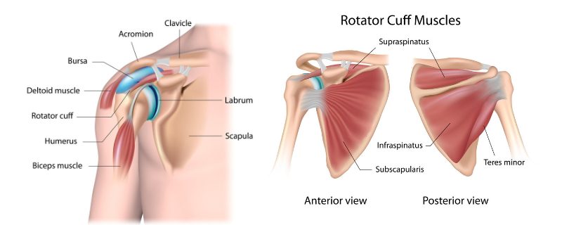
SHOULDER ANATOMY
The shoulder is a complex structure made up of three bones; the humerus (arm bone), the scapula (shoulder blade), and the clavicle (collar bone). At the top of the humerus is a large ball called the humeral head, which glides and rotates against a small, slightly curved part of the scapula called the glenoid (shoulder socket); the relatively small socket allows significant joint movement and flexibility. A firm, rubbery ring of tissue called the labrum is attached to the edge of the glenoid, and the humeral head and glenoid are covered in a smooth, gliding surface called articular cartilage (joint surface).
The bones of the shoulder form the glenohumeral joint (the true shoulder joint) and the acromioclavicular joint (the small joint between the acromial process of the scapula and the clavicle), and are joined together by ligaments (glenohumeral, coracoclavicular and acromioclavicular ligaments) and joint capsule.
The four rotator cuff muscles arise from the scapula and insert onto the humeral head, surrounding and supporting the glenohumeral joint and controlling fine shoulder movements. The strong chest wall muscles (deltoid, pectoralis major, lattisimus dorsi) and arm muscles (biceps, triceps) provide additional support and strength to the shoulder joint.
SHOULDER CONDITIONS
Dr. Botma treats the following conditions:
- Shoulder dislocation
- Shoulder instability
- Glenoid labral tears
- Rotator cuff tears – frequently asked questions
- Long head of biceps tendonitis
- Shoulder impingement
- Shoulder injuries in the throwing athlete
- SLAP tears (superior labrum from anterior to posterior tear)
- Acromioclavicular (AC) joint dislocation
- Acromioclavicular (AC) joint arthritis
- Frozen shoulder (adhesive capsulitis)
- Calcific tendonitis of the shoulder
- Suprascapular nerve compression
SHOULDER PROCEDURES
Dr. Botma performs the following shoulder procedures:
- Arthroscopic capsular plication
- Arthroscopic rotator cuff repair
- Biceps tenodesis
- Subacromial decompression surgery
- SLAP repair
- Acromioclavicular (AC) joint reconstruction
- Distal clavicle excision
- Frozen shoulder release
- Suprascapular nerve decompression
YOUR ELBOW
Your shoulder is the most flexible and mobile joint in your body, requiring a complicated interaction of bones, muscles, tendons and ligaments to function. Any of these structures can be damaged by injury or can slowly degenerate over time, causing pain, instability, stiffness or weakness.
Many shoulder problems do not require surgery and you may return to normal after a structured rehabilitation programme. If you do require shoulder surgery, Dr. Botmas will perform this using the most contemporary arthroscopic or minimally invasive techniques.
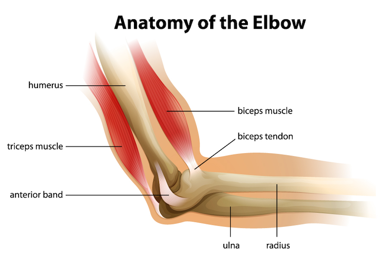
ELBOW ANATOMY
The elbow is a hinged joint that connects the humerus (upper arm bone) and the bones of the forearm (ulna and radius). Cartilage cushions the joint and allows the bones to slide smoothly against one another.
The medial collateral ligament on the inside of the elbow and the lateral collateral ligament on the outside of the elbow hold the bones together and provide stability. The annular ligament holds the head of the radius against the ulna.
Tendons attach muscle to bone. The biceps tendon attaches the biceps muscle at the front of the arm while the triceps tendon attaches the triceps muscle at the back of the arm. Most of the muscles are either attached to the lateral epicondyle, which is the bump on the outside of the elbow, or the medial epicondyle which is the bump on the inside of your elbow.
ELBOW CONDITIONS
Dr. Botma performs the following procedures:
- Golfer’s Elbow (medial epicondylitis)
- Tennis Elbow (lateral epicondylitis)
- Bursitis
- Elbow Osteoarthritis
- Cupital Tunnel Syndrome
- Elbow Dislocation
- Olecranon Bursitis
ELBOW PROCEDURES
Dr. Botma performs the following shoulder procedures:
- Elbow arthroscopy
- Arthroscopic Surgery For Elbow Bursitis
- Minimally Invasive Surgery For Rheumatoid Arthritis (RA)
- Elbow Surgery For Tennis Elbow
- Elbow Arthroscopy for Golfer’s Elbow
- Surgical Debridement for Osteochondritis Dissecans (OD)
YOUR HAND AND WRIST
The hand in the human body is made up of the wrist, palm, and fingers. The most flexible part of the human skeleton, the hand enables us to perform many of our daily activities. When our hand and wrist are not functioning properly, daily activities such as driving a car, bathing, and cooking can become impossible.
The hand’s complex anatomy consists of 27 bones, 27 joints, 34 muscles, over 100 ligaments and tendons, numerous blood vessels, nerves, and soft tissue.
HAND AND WRIST ANATOMY
- At the upper end of the wrist are the pisiform, triquetrum, lunate and scaphoid.
- On the lower side of the hand are the hamate, capitate, trapezoid and trapezium.
Within the hand are the:
- Metacarpals – the five bones in the middle part of the hand.
- Phalanges – the 14 bones that make up the fingers of each hand. Each finger has three phalanges (distal, middle and proximal) while the thumb has two.
Where two bones meet are the joints. Each finger has three joints:
- At the bases is the metacarpophalangeal joint.
- In the middle is the proximal interphalangeal joint.
- At the end of the fingers is the distal interphalangeal joint.
Cartilage covers the end of the bones in the joint, lubricated by synovial fluid. This allows them to glide smoothly over one another as the joints move.
Ligaments connect the bones, helping to support them and provide stability. The ligaments in the hand include:
- Collateral ligaments on either side of the finger and thumb joints.
- Volar plate ligaments.
- Radial and ulnar collateral ligaments.
- Volar radiocarpal ligaments.
- Dorsal radiocarpal ligaments.
- Ulnocarpal and radioulnar ligaments.
Tendons connect muscles to bone. Among the tendons in the hand are the superficialis, profundus, extensor, flexor, extensor pollicis brevis and abductor pollicis longus.